Horse Anatomy: focus on the rear limbs
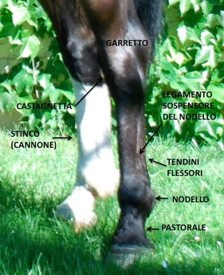
In this Equestrian World piece dedicated to horse anatomy, the focus will be on the posterior limbs of a horse.
The hind limbs are connected to the pelvis through the femoral hip joint, the pelvis is in turn connected to the spine through the sacroiliac joint.
The joint is surrounded by large areas of muscle mass that propel the hind limbs. The connection between the hind limb muscles and front limbs occurs through the large back muscle that runs through the spine from the cervical vertebrae to the pelvis, with the help of a ligament that supports the head.
Tendons are similar to ropes, they tie into a fibrous tendon and a synovial fluid lubricates and prevents friction, protecting against wear and tear of the tissues. The ligaments have no similar protection, and their function is precisely to “bind“ and connect muscle tissues.
Muscles, tendons and ligaments, thanks to their elasticity create in the first phase of the movement thrust forces, while in the second phase the pressure lessens.
SOME REAR MUSCLES AND TENDONS ARE MORE PRONE TO INJURY OR DISEASE THAN OTHERS.
The forearm and the related tendons are a source of injury.
1 ) SHORT AND BIG MUSCLE EXTENDER (forearm) . Their tendons are inserted on the front of the carpus.
2 ) BIG MUSCLE EXTENDER (phalange). Its tendon extends on the front face of the limb.
3 ) SURFACE AND DEEP MUSCLE FLEXORS (phalange) . The tendons are at the rear of the limb.
Between the deep tendon flexor and metacarpal-metatarsal there is a big ligament which half-way up the shin divides into two branches for support.
The most common injuries in the back legs are often found in the quadriceps and ankle muscle. In the next Equestrian World piece we will look at nomenclature.
from Dr. Vittorio Meschia’s book published by Horse S.r.l


