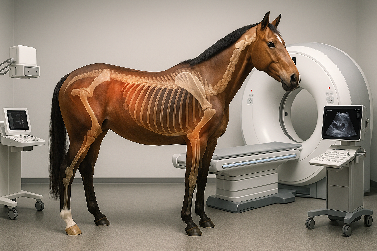
The evolution of Diagnostic imaging in equine medicine: from X-Rays to molecular scanning

When Diagnosis Was Blind
At the beginning of the 20th century, veterinary medicine relied almost entirely on human senses: sight, touch, and hearing.
Lameness, swelling, mysterious pain — all were interpreted through careful observation and palpation.
There was no way to “see” inside a horse’s body.
Veterinary science was more an art based on experience than a discipline grounded in objective evidence.
Then, at the end of the 19th century, science opened a new window into the invisible.
1895: The Discovery of X-Rays – The Birth of Radiography
In 1895, Wilhelm Conrad Röntgen, experimenting with electrical discharge tubes, accidentally discovered a form of invisible radiation that could penetrate solid matter: X-rays.
When captured on a photographic plate, these rays revealed the internal structure of living beings — humans and animals alike.
Veterinarians quickly recognized the potential.
Early equine radiographs were far from perfect: machines were massive, exposure times were long, and images were often blurred.
Yet even through those primitive images, fractures, bone deformities, and joint abnormalities became visible without the need for surgery.
It was the beginning of a revolution.
How does radiography work?
A beam of X-rays passes through the body and is absorbed differently by various tissues.
- Bones, being very dense, absorb most of the rays and appear white on the image.
- Soft tissues, less dense, allow more rays to pass through, appearing darker.
As technology advanced, portable X-ray machines were developed, making radiography a practical daily tool in equine clinics and racetracks.
1950–1970: Modern Radiography – Safer, Sharper Images
After World War II, advancements in X-ray tubes and film technology dramatically improved image quality.
Radiography became a cornerstone of equine diagnostics, particularly for sport and racehorses.
However, X-rays inherently provided clear views of hard tissues like bones but revealed little about soft tissues such as tendons, ligaments, and muscles.
A new technology was needed to bridge that gap.
The 1970s: Ultrasound – Listening to the Body
In the 1970s, a groundbreaking innovation arrived: ultrasound.
Rather than using radiation, ultrasound employed high-frequency sound waves.
How does ultrasound work?
A handheld probe, placed against the skin, emits sound waves into the body.
When these waves encounter different tissues, they reflect back with varying intensity.
The echoes are captured and converted into real-time images by a computer.
Ultrasound became crucial for:
- Visualizing tendons and ligaments, essential for equine athletes.
- Examining muscles and joint capsules.
- Monitoring pregnancies and fetal development.
Its non-invasive, painless, and field-portable nature made it an indispensable tool for modern veterinary practice.
The 1980s: CT Scans – Slicing the Body into Images
In the 1980s, Computed Tomography (CT) added a whole new dimension to veterinary imaging.
How does CT work?
An X-ray source rotates around the horse while detectors measure the radiation passing through the body from different angles.
A computer reconstructs these measurements into highly detailed cross-sectional images — like slices through the body.
CT is particularly useful for:
- Detecting complex fractures.
- Studying the skull, sinuses, and spine.
- Planning intricate surgical interventions.
To obtain clear images, horses typically need to be sedated or anesthetized during CT scans.
The 1990s: MRI – Mapping the Soft Tissues
Following CT, Magnetic Resonance Imaging (MRI) took the visualization of soft tissues to an unprecedented level.
How does MRI work?
MRI uses a strong magnetic field to align hydrogen atoms in the body’s tissues.
Radiofrequency pulses disturb this alignment, and as the atoms return to their original positions, they emit signals captured and processed into detailed images.
MRI allows veterinarians to:
- Visualize tendons, ligaments, cartilage, menisci, and bone marrow with extraordinary precision.
- Diagnose subtle injuries invisible on X-rays or CT scans.
- Investigate complex joint diseases and unexplained lameness.
Today, standing MRI systems enable horses to be scanned while awake, reducing the risks associated with anesthesia.
Functional Imaging – Watching Biology in Action
At the dawn of the 21st century, imaging evolved from capturing structure to observing function.
With bone scintigraphy, a small amount of radioactive tracer (typically Technetium-99m) is injected into the bloodstream.
The tracer accumulates in areas of high bone activity, such as inflammation or microfractures, and is detected by a gamma camera.
Positron Emission Tomography (PET) takes this even further, allowing real-time observation of cellular metabolism, enabling early detection of:
- Stress injuries.
- Bone infections.
- Tumors.
These functional imaging techniques are now vital tools for a new era of precision equine medicine.
The Present and the Future: A Journey Still Unfolding
Today, diagnostic imaging in horses is no longer a luxury; it is an essential element of modern veterinary practice.
Radiography, ultrasound, CT, MRI, scintigraphy, and PET work in concert to provide a complete, nuanced view of equine health.
Each technique offers a different perspective, helping veterinarians diagnose more accurately, treat more effectively, and protect the future of these magnificent animals.
The journey that began with a faint, flickering shadow on a photographic plate continues to unfold — and its next steps promise to be even more extraordinary.
Alessia Niccolucci
© Rights Reserved.


