Veterinary Science: the structure of a horse’s hoof (part 1).
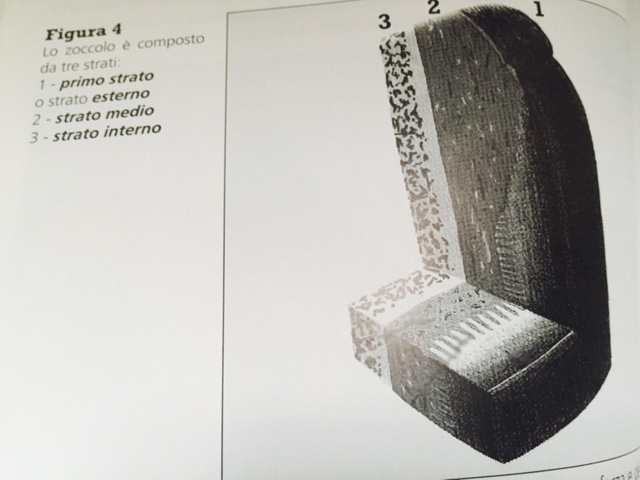
The microscopic structure of a horse’s foot is one of the great miracles of nature. This brief description in this Equestrian World piece will help to understand how the internal structure of the base is designed to achieve high levels of strength and flexibility to support the third phalanx, protecting it from the shock of the impact with the ground. A reminder that when galloping there is a phase in which the full weight of the horse is on one foot.
COMPOSITION OF THE HOOF
The hoof is made up of three layers: the first layer (outer layer) which we can see with the naked eye and is known as the nail.
The middle layer forms the biggest part of the wall and is a non-pigmented material of microscopic thorny small tubes surrounded by a tissue called the inter-tubular nail.
The thorny small tubes are responsible for the strength and elasticity of the hoof, and within them, there are cells that absorb the water that then allow the small tubes to flex and compress.
The internal layer consists of 600 or more tissues and are extremely intelligent combining both power and elasticity.
From “La zoppia del cavallo, conoscere per capire / the lameness of the horse, learn to understand.“ Dr. Vittorio Meschia, Horse srl.
All rights reserved ©


.png)


